Discover and read the best of Twitter Threads about #ECG
Most recents (24)

1) Welcome to Part 1 of a new #accredited #tweetorial in our series of educational programs on #hypertrophic #cardiomyopathy #HCM. Previous programs, still available for 🆓CE/#CME, are at cardiometabolic-ce.com/category/hcm/.
Now you can earn another 1.5hr credit by following ALL of this 🧵!
Now you can earn another 1.5hr credit by following ALL of this 🧵!
2) Our expert author is Sergio Kaiser MD PhD FACC FESC 🇧🇷🇮🇱 @pabeda1, cardiologist 🫀, Professor 🎓 of #InternalMedicine, Rio de Janeiro State University. He brings the general cardiologist's perspective to our #HCM discussions. Read and learn!
#FOAMed #CardioTwitter
#FOAMed #CardioTwitter

3) This program is supported by an unrestricted educational grant from Bristol Myers Squibb. Statement of accreditation and faculty disclosures at cardiometabolic-ce.com/disclosures/. Credit for #physicians #nursepractitioners #physicianassociates #nurses #pharmacists from @academiccme.

When your ER colleague calls you to have a quick look on an #ecg … the 28 yo pat reports frequent episodes of palpitations since childhood that he can usually terminate with deep breathing. #Epeeps @EPeeps_Bot #CardioTwitter know what’s going on. Do’s and Don’t’s? twitter.com/i/web/status/1…
10 min after giving 2mg/kg body weight Flecainid iv. twitter.com/i/web/status/1…
And 2 hours later


1/ Let’s talk about focal atrial tachycardia #CardioTwitter.
🧵👇 by @ekgdx
#ekgdx #ecg #ekg #MedTwitter #medicine #medstudents #basic #education #graphic
🧵👇 by @ekgdx
#ekgdx #ecg #ekg #MedTwitter #medicine #medstudents #basic #education #graphic

2/ Focal atrial tachycardia is characterized by at least three or more consecutive ectopic P waves with similar morphology, usually arising from a single ectopic focus.
#ekgdx
#ekgdx
3/ Criteria
✅ ≥3 consecutive similar ectopic P waves (usually inverted in inferior leads).
✅ Atrial rate >100 bpm.
✅ QRS usually narrow unless pre-existing BBB or aberrant conduction.
#ekgdx
✅ ≥3 consecutive similar ectopic P waves (usually inverted in inferior leads).
✅ Atrial rate >100 bpm.
✅ QRS usually narrow unless pre-existing BBB or aberrant conduction.
#ekgdx

A young man was found down in cardiac arrest. 911 was called and paramedics achieved ROSC en route prior to arrival in the ER. No further history was available. This #ECG was recorded.
What’s the diagnosis?
What’s the diagnosis?

If you’re a new follower I always post the answer with explanation the next day. If you’re new to my account—follow me if you want to learn about medical emergencies
Answer:

AV dissociation, capture beats and fusion beats are all hallmarks of ventricular tachycardia
A simple understanding 👇
#CardioTwitter #Cardiology #Medtwitter
A simple understanding 👇
#CardioTwitter #Cardiology #Medtwitter
What’s a “capture beat” ?
During ventricular tachycardia, sometimes the SA nodal firing takes control of independent ventricular depolarization for brief moment, as this happens, “CAPTURE BEATS” appear. They are nothing but a brief moment of normal looking P-QRS pattern
During ventricular tachycardia, sometimes the SA nodal firing takes control of independent ventricular depolarization for brief moment, as this happens, “CAPTURE BEATS” appear. They are nothing but a brief moment of normal looking P-QRS pattern
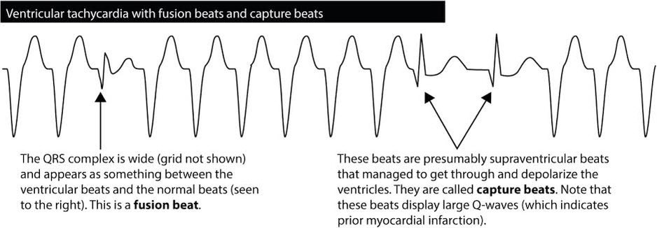
Whats a “fusion beat” ?
During ventricular tachycardia, sometimes the SA nodal impulse and the independent ventricular impulse combine together to create a mix looking P-QRS pattern called fusion beats.
#ECG @EPeeps_Bot #ecglearning #CardiacArrest
During ventricular tachycardia, sometimes the SA nodal impulse and the independent ventricular impulse combine together to create a mix looking P-QRS pattern called fusion beats.
#ECG @EPeeps_Bot #ecglearning #CardiacArrest


1/ Let’s talk about T waves #CardioTwitter
The T wave represents typically ventricular repolarization. It is the most labile wave on the EKG surface.
Normal T wave
✅ Morphology: Asymmetric.
✅ Amplitude: ≤6 mm in limb leads and ≤10 mm in precordial leads.
#MedTwitter
The T wave represents typically ventricular repolarization. It is the most labile wave on the EKG surface.
Normal T wave
✅ Morphology: Asymmetric.
✅ Amplitude: ≤6 mm in limb leads and ≤10 mm in precordial leads.
#MedTwitter

2/ Tall upright T wave
Tall upright T waves are usually characterized by tall and peaked shape.
✅ Amplitude: >6 mm in limb leads and >10 mm in precordial leads.
Causes: Hyperkalemia, hyperacute MI, normal variant, prinzmetal angina, aortic stenosis, LVH, RVH, others.
#ecg
Tall upright T waves are usually characterized by tall and peaked shape.
✅ Amplitude: >6 mm in limb leads and >10 mm in precordial leads.
Causes: Hyperkalemia, hyperacute MI, normal variant, prinzmetal angina, aortic stenosis, LVH, RVH, others.
#ecg

3/ Notched T wave
Possible causes: May be caused by morphological changes in the cardiomyocytes' action potential waveforms. Another causes include: Drugs (such as Dofetilide, Quinidine, Ranolazine, Verapamil), long QT syndrome, athletes, others.
#MedTwitter #MedStudentTwitter
Possible causes: May be caused by morphological changes in the cardiomyocytes' action potential waveforms. Another causes include: Drugs (such as Dofetilide, Quinidine, Ranolazine, Verapamil), long QT syndrome, athletes, others.
#MedTwitter #MedStudentTwitter


Are you a #juniordoctor or #medstudent?
Here's 10 great FREE modules to help get you started on the wards!
#meded #medschool #tipsfornewdocs #juniordocs #FOAMed #medtwitter #medstudenttwitter #juniordoctors #medstudents
Here's 10 great FREE modules to help get you started on the wards!
#meded #medschool #tipsfornewdocs #juniordocs #FOAMed #medtwitter #medstudenttwitter #juniordoctors #medstudents
Occasionally you'll need to perform sterile procedures. Make sure you prepare the best you can
osler.app.link/CztXIRjyntb
osler.app.link/CztXIRjyntb

Providing basic life support is a core skill for all healthcare staff
osler.app.link/u4fe8uqyntb
#basiclifesupport #bls #FOAMresus #resuscitation
osler.app.link/u4fe8uqyntb
#basiclifesupport #bls #FOAMresus #resuscitation

1/ Let’s talk about the ST segment #CardioTwitter.
ST segment normally represents the interval between ventricular depolarization and repolarization.
Normal ST
✅ Usually isolectric or may vary from 0.5 mm below to 1 mm above isolectric line in L leads.
#ekgdx #Medicine
ST segment normally represents the interval between ventricular depolarization and repolarization.
Normal ST
✅ Usually isolectric or may vary from 0.5 mm below to 1 mm above isolectric line in L leads.
#ekgdx #Medicine

2/ ST elevation (STE)
ST changes suggesting myocardial injury:
✅ New STE ≥1 mm in all leads other than V2 or V3.
✅ New STE in V2-V3 ≥2 mm in men older than 40 years old and ≥2.5 mm in men younger than 40 years old or ≥1.5 mm in women.
#ekgdx #Medstudent #MedTwitter
ST changes suggesting myocardial injury:
✅ New STE ≥1 mm in all leads other than V2 or V3.
✅ New STE in V2-V3 ≥2 mm in men older than 40 years old and ≥2.5 mm in men younger than 40 years old or ≥1.5 mm in women.
#ekgdx #Medstudent #MedTwitter

3/ Types of ST segment elevation include:
✅ Convex Upward (previous pic)
✅ Horizontal (this one)
✅ Concave Upward (see next)
✅ Obliquely Straight (see next)
#ekgdx #st #MedTwitter #ecg #ekg #CardioTwitter #Medstudent
✅ Convex Upward (previous pic)
✅ Horizontal (this one)
✅ Concave Upward (see next)
✅ Obliquely Straight (see next)
#ekgdx #st #MedTwitter #ecg #ekg #CardioTwitter #Medstudent


Three #cardiology cases with diagnostic ECGs in our resus room today and some learning points for emergency clinicians
#ecg #ekg
#ecg #ekg
1. Sudden onset palpitations
ECG shows regular narrow complex tachycardia with rate around 140
ECG shows regular narrow complex tachycardia with rate around 140

We suspected this was atrial flutter
Rather than subject a patient to the horror of iv adenosine (which only reveals flutter - it can’t convert it), we moved the ECG limb leads around to get a ‘Lewis lead’ which better shows atrial activity
(See litfl.com/lewis-lead-s5-… )
Rather than subject a patient to the horror of iv adenosine (which only reveals flutter - it can’t convert it), we moved the ECG limb leads around to get a ‘Lewis lead’ which better shows atrial activity
(See litfl.com/lewis-lead-s5-… )

1/ Let’s talk about PR Interval - Segment #CardioTwitter.
The PR interval represents the time between the onset of atrial depolarization and the onset of ventricular depolarization and reflects conduction through the AV node.
🧵by @ekgdx
The PR interval represents the time between the onset of atrial depolarization and the onset of ventricular depolarization and reflects conduction through the AV node.
🧵by @ekgdx

2/ PR Interval
Criteria
✅ Normal PR interval: 0.12 - 0.20 sec.
✅ Prolonged PR interval: >0.20 sec.
✅ Short PR interval: <0.12 sec.
The term “PQ interval” is preferred by some EKG lover because it is the period actually measured unless the Q wave is absent.
#PR #interval
Criteria
✅ Normal PR interval: 0.12 - 0.20 sec.
✅ Prolonged PR interval: >0.20 sec.
✅ Short PR interval: <0.12 sec.
The term “PQ interval” is preferred by some EKG lover because it is the period actually measured unless the Q wave is absent.
#PR #interval
3/ The PR segment is the segment between the end of the P wave and the start of the QRS complex.
Criteria
✅ Normal PR segment: Usually isolectric.
✅ PR segment elevation: ≥0.5 mm.
PR segment elevation causes: Atrial ischaemia/infarction, myopericarditis, PE, others.
#ekgdx
Criteria
✅ Normal PR segment: Usually isolectric.
✅ PR segment elevation: ≥0.5 mm.
PR segment elevation causes: Atrial ischaemia/infarction, myopericarditis, PE, others.
#ekgdx


🆘59 year male smoker
k/c/o COPD, DM, HTN with DCMP (EF=20%) palpitations and dizziness x 2 days
O/E : B/L crepts +, rest
ECG👇
#Medtwitter #MedEd2022 #ECG #CardioEd
k/c/o COPD, DM, HTN with DCMP (EF=20%) palpitations and dizziness x 2 days
O/E : B/L crepts +, rest
ECG👇
#Medtwitter #MedEd2022 #ECG #CardioEd

What is the likely rhythm ?
Answer - 𝘼𝙩𝙧𝙞𝙖𝙡 𝙁𝙞𝙗𝙧𝙞𝙡𝙡𝙖𝙩𝙞𝙤𝙣
📍Absent discrete P-waves
📍Fibrillatory waves (f-waves)
📍Irregularly irregular QRS complexes
📍Absent discrete P-waves
📍Fibrillatory waves (f-waves)
📍Irregularly irregular QRS complexes

1/ Let’s talk about P waves #CardioTwitter.
The P wave is the first positive deflection on the EKG and represents atrial depolarization. The first half represents right atrial depolarization and the second half represents left atrial depolarization.
By @ekgdx
The P wave is the first positive deflection on the EKG and represents atrial depolarization. The first half represents right atrial depolarization and the second half represents left atrial depolarization.
By @ekgdx

2/ Normal P wave
Criteria
✅ Axis: 0° to +75°.
✅ Amplitude (L leads): <2.5 mm.
✅ Amplitude (P leads): <1.5 mm.
✅ Duration: 0.08 - 0.11 sec.
✅ Morphology: Upright in I, II, aVF and inverted in aVR.
#Pwaves #ecg #ekg #ekgdx #medicine #MedTwitter #MedStudentTwitter #basic
Criteria
✅ Axis: 0° to +75°.
✅ Amplitude (L leads): <2.5 mm.
✅ Amplitude (P leads): <1.5 mm.
✅ Duration: 0.08 - 0.11 sec.
✅ Morphology: Upright in I, II, aVF and inverted in aVR.
#Pwaves #ecg #ekg #ekgdx #medicine #MedTwitter #MedStudentTwitter #basic
3/ Peaked P wave
The morphology is peaked with amplitude ≥2.5 mm, usually in II, III and aVF.
The peaked P wave is a typical characteristic of right atrial abnormality/enlargement.
Causes: PE, COPD, congenital heart disease, pulmonary hypertension, normal variant, others.
The morphology is peaked with amplitude ≥2.5 mm, usually in II, III and aVF.
The peaked P wave is a typical characteristic of right atrial abnormality/enlargement.
Causes: PE, COPD, congenital heart disease, pulmonary hypertension, normal variant, others.

Acute pulmonary embolism (PE) is one of the most serious form of venous thromboembolism. The clinical presentation of PE is variable and often nonspecific making the diagnosis challenging.
1/
#CardioTwitter
1/
#CardioTwitter

Criteria
✅ Sinus tachycardia (most common).
✅ S1Q3T3 pattern (may be present up to 30% of cases).
✅ Simultaneous T wave inversions in the inferior leads and right precordial leads can be seen.
✅ Right axis deviation.
✅ RBBB (complete or incomplete).
2/
#MedTwitter #ecg
✅ Sinus tachycardia (most common).
✅ S1Q3T3 pattern (may be present up to 30% of cases).
✅ Simultaneous T wave inversions in the inferior leads and right precordial leads can be seen.
✅ Right axis deviation.
✅ RBBB (complete or incomplete).
2/
#MedTwitter #ecg

Here’s a very important #ECG of a 60-year-old lady who presented with chest pain and shortness of breath that began while walking her dog. She was also COVID+
What’s the diagnosis?
What’s the diagnosis?

If you’re a new follower I always post the answer with explanation the next day. If you’re new to my account—follow me if you want to learn about medical emergencies
Here’s a very important #ECG that was recorded in a 50-year-old lady shortly before she suddenly went into cardiac arrest
What’s the diagnosis?
What’s the diagnosis?

If you’re a new follower I always post the answer with explanation the next day. If you’re new to my account—follow me if you want to learn about medical emergencies


Its WPW pattern --> Derpak Sir has provided a methology to localize the Kent bundle --> I can't call it WPW syndrome for there is no hx of pre excited NCT.
*Deepak Sir

Please retweet and share if you support my and others' vaccine injury recoveries.

My fiancé and I were supposed to get married in July 2022. Given the first 2 vaccine mandates, we figured that we had no choice but to keep on taking the boosters in order to be able to travel for our honeymoon / have liberties to go to stores etc.
We decided to take the 3rd @moderna_tx booster anticipating that perhaps it would also be mandated by the #Canadian #government.
Here’s the #ECG of a 68 year old man who was rushed to the ER by paramedics
BP: 80/40
HR: 150
RR: 35
SPO2: 95%
What’s the diagnosis?
BP: 80/40
HR: 150
RR: 35
SPO2: 95%
What’s the diagnosis?

Answer: HYPERKALEMIA
There’s a wide complex tachycardia with RBBB morphology. There are features here concerning for several life-threatening diagnoses including: V-Tach, Pulmonary Embolism, Acute Coronary Occlusion. But ALL these changes resolved with empiric ↑K+ treatment…
There’s a wide complex tachycardia with RBBB morphology. There are features here concerning for several life-threatening diagnoses including: V-Tach, Pulmonary Embolism, Acute Coronary Occlusion. But ALL these changes resolved with empiric ↑K+ treatment…
The QRS width narrowed right before our eyes. His vital signs improved dramatically. This was the repeat ECG recorded just 20 minutes later. Lead V1 is artifact but otherwise you can see that all the extreme changes have now resolved, and only a hint of peaked T waves remain…


🧵 1/7 Ever wondered why the Osborn wave looks the way it does? Stay with me during my newest #tweetorial. A thread 🧵1/7
#cardiotwitter #EPeeps #CardioEd #MedTwitter @TRassafMD @YoungDgk @DGK_org @YoungDZHK @AaronGoodman33 @Steph_Achenbach @fuzzymittens @AvrahamCooperMD
#cardiotwitter #EPeeps #CardioEd #MedTwitter @TRassafMD @YoungDgk @DGK_org @YoungDZHK @AaronGoodman33 @Steph_Achenbach @fuzzymittens @AvrahamCooperMD

2/7 History
First described in 1953 by Osborn (camel-hump sign) upon #hypothermia in dogs. Upon systemic analysis similar #ECG patterns have been described in
➡️ hypercalcemia
➡️ brain injury
➡️ SAB
➡️ vasospastic angina / ischemia
First described in 1953 by Osborn (camel-hump sign) upon #hypothermia in dogs. Upon systemic analysis similar #ECG patterns have been described in
➡️ hypercalcemia
➡️ brain injury
➡️ SAB
➡️ vasospastic angina / ischemia
3/7 Emslie-Smith et al showed that Osborn waves manifested more in epicardial than endocardial leads. Others finally showed that 4-aminopyridine sensitive transient outward current (Ito) is responsible and predominantly located in epicardium. ⬇️ heart rate led to ⬆️ Ito current
Here’s an incredible #ECG recorded in a 60 year old man with chest pain and palpitations
What’s the diagnosis?
What’s the diagnosis?

If you’re a new follower I always post the answer with explanation the next day. If you’re new to my account—follow me if you want to learn about Emergency ECGs

My 1st symptoms of #COVID were 7 weeks ago tomorrow.
Yesterday I went for an #ECG b/c I'm having heart symptoms that weren't there before #COVID19.
I'm also having issues with my blood sugar which weren't there before #Covid_19
The technician said he's doing several ECG's ++
Yesterday I went for an #ECG b/c I'm having heart symptoms that weren't there before #COVID19.
I'm also having issues with my blood sugar which weren't there before #Covid_19
The technician said he's doing several ECG's ++
daily on people who are having heart complications due to #COVID19.
I'm on leave from my job. And now I'm in need of health care I wasn't before. I'm scared of what is happening inside my body and brain.
@TimHoustonNS got rid of all the means that protected the #NovaScotia ++
I'm on leave from my job. And now I'm in need of health care I wasn't before. I'm scared of what is happening inside my body and brain.
@TimHoustonNS got rid of all the means that protected the #NovaScotia ++
population. Our health care and workers are crumbling under the weight of it all.
Businesses are closing. Housing is a mess. The climate is in shambles.
What are we going to do?
#nspoli #covid19NS #CovidIsNotOver #COVID19 #COVID
Businesses are closing. Housing is a mess. The climate is in shambles.
What are we going to do?
#nspoli #covid19NS #CovidIsNotOver #COVID19 #COVID
Here’s an important #ECG that was recorded in a 50-year-old man just 15 minutes before he suddenly went into pulseless V-Tach
What’s the diagnosis?
What’s the diagnosis?

This was an unbelievable case. If you’re a new follower I always post the answer with explanation the next day. If you’re new to my account—follow me if you want to learn about Emergency ECGs

Hey #OhGottPJ - Ihr seid alleine in der ZNA und seht diesen Monitorausschnitt - was passiert da? Und was jetzt? :D
#ekg #ecg #cardiotwitter
#ekg #ecg #cardiotwitter

Man sieht irgendwas, was zu einer Breitkomplextachykardie umspringt in der Monitorüberwachung.
1. Wir sind uns ja relativ einig es handelt sich um einen potentiellen Notfall also:
In case of emergency, take your own pulse first!
1. Wir sind uns ja relativ einig es handelt sich um einen potentiellen Notfall also:
In case of emergency, take your own pulse first!
2. Hilfe rufen (alleine ist blöd)
Here’s a great #ECG of a 60 year old man with chest pain and shortness of breath. This is a fantastic case—with a twist!
What’s the diagnosis?
What’s the diagnosis?

If you’re a new follower I always post the answer with explanation the next day. If you’re new to my account, follow me if you want to learn about Emergency ECGs
Here’s a video I made breaking down this rare and fascinating case of a 60 year old man with chest pain and shortness of breath
#FOAMed
#FOAMed
