Discover and read the best of Twitter Threads about #microscopy
Most recents (24)

How do you image the large and the small at the same time? We developed new 🔬 technology to image centimeter-scale specimens - including whole mouse brains 🧠 - with diffraction-limited resolution and without sectioning. #mesoscale #imaging
biorxiv.org/content/10.110…
🧵 (1/n)
biorxiv.org/content/10.110…
🧵 (1/n)
ExA-SPIM is a technology based on innovations in (1) tissue processing and (2) large-scale microscopy 🔬. Our team developed new methods for expansion for microscopy (ExM) of large tissue volumes. #teamscience #expansionmicroscopy
protocols.io/view/whole-mou…
🧵 (2/n)
protocols.io/view/whole-mou…
🧵 (2/n)
By combining ExM with a new SPIM system based on technologies from electronics metrology, ExA-SPIM pushes past the volumetric imaging barrier 🚧 that constrains previous imaging approaches. (inspired by recent plot from @Daetwyler_St, @RetoPaul) #lightsheet #microscopy
🧵 (3/n)
🧵 (3/n)

What are we looking at?
Is this a tubular object?
-Case H&E 1108
Male 65 years
Warty lesion on face
Diagnosis:
Squamous carcinoma
(Via Virtual Pathology Slide Library)
#dermatology #Microscopy #glochid
Is this a tubular object?
-Case H&E 1108
Male 65 years
Warty lesion on face
Diagnosis:
Squamous carcinoma
(Via Virtual Pathology Slide Library)
#dermatology #Microscopy #glochid


A section of a tube is an ellipsoid, which can be a circle when the section is perpendicular to the tube.


#quantum is here! Why not collect #fluorescence #photon-by-photon in your laser-scanning #microscope? We developed the #BrightEyes_TTM, an #opensource data acquisition system able to do it!
nature.com/articles/s4146…
@IITalk @ERC_Research @RNA_iit @MSCActions #BrightEyes_ERC
(1/5)
nature.com/articles/s4146…
@IITalk @ERC_Research @RNA_iit @MSCActions #BrightEyes_ERC
(1/5)
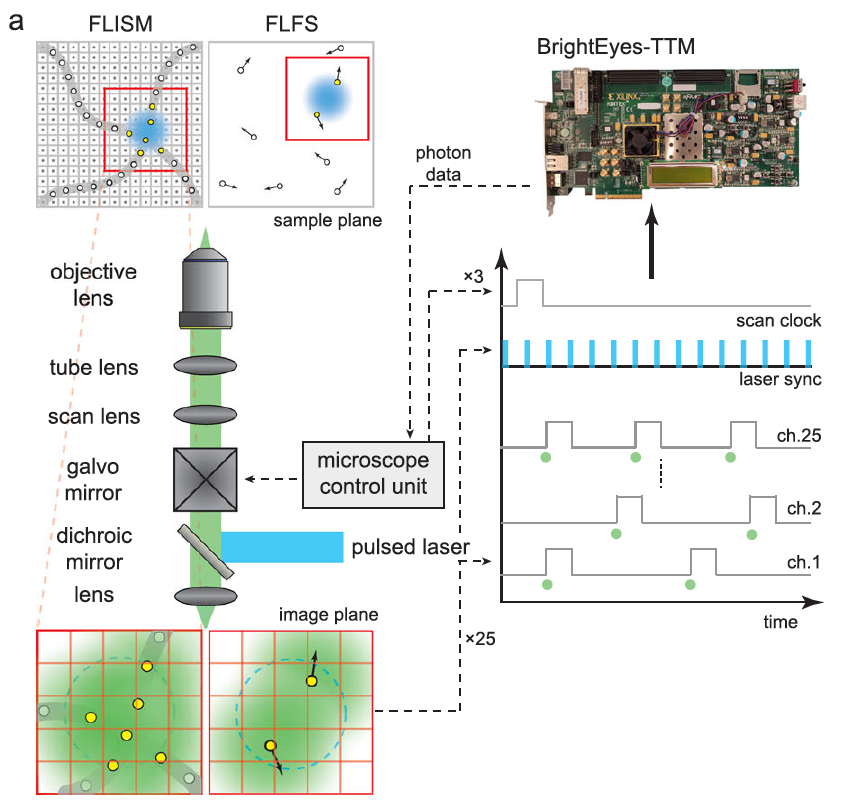
Our time-tagging #DAQ module can collect single-photon signals from 25 independent channels and reference signals from 3 channels. We showed how this module combined with a #SPAD array allows us to perform #FLISM and #FLFS. (2/5)
#imagescanningmicroscopy #fluorescence #lifetime
#imagescanningmicroscopy #fluorescence #lifetime
We characterized the #BrightEyes_TTM. We can reach a single-shot precision of 30 ps, 4 ns dead-time, and an "infinite" range in all channels, with a total flux of up to 125 MHz. (3/5)
#TCSPC #antibunching #quantum #microscopy #FPGA
#coding #LIDAR
#TCSPC #antibunching #quantum #microscopy #FPGA
#coding #LIDAR


Labeling bio images for AI just got 10x faster🏎. Today, we’re releasing Biodock Autolabel, and it’s kind of magical. We trained it on thousands of bio images so you can use it without training. (1/6)
@BiodockAI #microscopy #pathology #biology
@BiodockAI #microscopy #pathology #biology
Labeling objects is one of the most painful parts of training AI, especially in bio. Clicking 40 times or dragging the pen tool to define an ROI for hundreds of objects takes a very long time. And yet precise labels are so important for AI accuracy. (2/6)
Now, with Autolabel (powered by AI), this becomes two clicks (or one drag!). Our model takes your defined box and generates what it thinks is the precise object polygon label. For most biological objects, it just works, and it’s getting more and more accurate over time. (3/6)

Don't have access to a fancy Airyscan or STED?
Do have access to a good ole fashion confocal?
You can still do super resolution imaging!🧵#microscopy
Do have access to a good ole fashion confocal?
You can still do super resolution imaging!🧵#microscopy
All you need are good microscopy and digital imaging fundamentals. Everything you need to know can be found here: ibiology.org/online-biology… /2
Digital images are made up of spatially defined boxes called 'pixels'. Images are collections of pixels assigned color shades from a lookup table. /3


“Cryo-ExM” or “how to see (almost) everything you ever dreamt of”
Want to know more ?!? Check our story out with @CentrioleLab @nikolai_klena in @naturemethods: nature.com/articles/s4159… which combines cryo-fixation/Freeze substitution with #expansion #microscopy (#UExM).
Want to know more ?!? Check our story out with @CentrioleLab @nikolai_klena in @naturemethods: nature.com/articles/s4159… which combines cryo-fixation/Freeze substitution with #expansion #microscopy (#UExM).

1/Cryo-fixation, as demonstrated in electron microscopy, outperformed chemical fixations on subcellular structure such as the endoplasmic reticulum

2/Cryo-fixation circumvent the artifact of chemical fixations such as loss of antigenicity as seen with the mitochondrial outer membrane marker TOMM20


Daily Bookmarks to GAVNet 10/31/2021 greeneracresvaluenetwork.wordpress.com/2021/10/31/dai…
Adrienne LaFrance on Twitter and TreadReader
threadreaderapp.com/thread/1452591…
#facebook #transparency #profit #MoralCompass #manipulation
threadreaderapp.com/thread/1452591…
#facebook #transparency #profit #MoralCompass #manipulation
SpaceX needs to tame toilet trouble before weekend launch
phys.org/news/2021-10-s…
#SpaceX #nasa #ToiletLeaks #repairs
phys.org/news/2021-10-s…
#SpaceX #nasa #ToiletLeaks #repairs

It’s exciting to finally share and discuss our study on additive cytotoxicity, a crowd-based killing mechanism of cytotoxic T cells (CTL) to target cancer!
Highlights in the thread, free access @NatureComms here: nature.com/articles/s4146…
1/17
#immunology #microscopy
Highlights in the thread, free access @NatureComms here: nature.com/articles/s4146…
1/17
#immunology #microscopy
Initially, we aimed to study modulators of CTL serial killing capacity. But live-cell imaging of various mouse and human model systems, incl. OT1 CTL targeting OVA-expressing cancer cells surprisingly showed, that CTL are pretty bad killers and most contacts were non-lethal. 2/
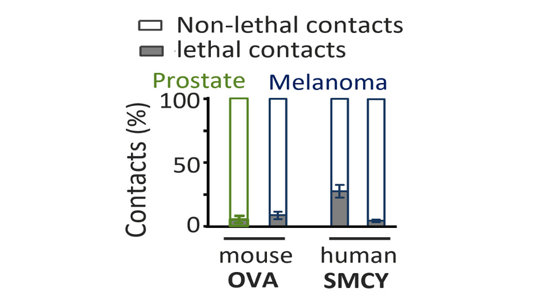
We doubted our ability to culture cytotoxic T cells.... but in co-cultures with fibroblast-like cells, the same OT1 CTLs proved to be efficient serial killers. 3/

We are pleased to announce that our #ZeroCostDL4Mic work is now published!
nature.com/articles/s4146…
And our @github
github.com/HenriquesLab/Z…
#DeepLearning and #Microscopy for all!
For the occasion, I’ve put a small Tweetorial together to explain what this is all about!
Thread 0/7
nature.com/articles/s4146…
And our @github
github.com/HenriquesLab/Z…
#DeepLearning and #Microscopy for all!
For the occasion, I’ve put a small Tweetorial together to explain what this is all about!
Thread 0/7

Thread 1/7
You can implement Deep Learning for microscopy in several ways:
1- Using local resources (fast GPUs)
2- Using cloud-computing
3- Using pre-trained models
We think that (1) is uncommon, and that (3) can be unreliable. So we do (2)!
You can implement Deep Learning for microscopy in several ways:
1- Using local resources (fast GPUs)
2- Using cloud-computing
3- Using pre-trained models
We think that (1) is uncommon, and that (3) can be unreliable. So we do (2)!

Thread 2/7
We built a platform using @GoogleColab and @ProjectJupyter notebooks that can perform training, quality control and prediction, all on the cloud and for free!
All of this dedicated to #microscopy !
We built a platform using @GoogleColab and @ProjectJupyter notebooks that can perform training, quality control and prediction, all on the cloud and for free!
All of this dedicated to #microscopy !


Hola! my name is Ale (they/she) and I'm taking part in the #GlobalScienceShow
Today I'll be showing you some of my works about my exploration of the microbial world with drawings! 🦠
Check @GlobalSciShow to follow the whole show🤩
#scicomm #divulgarciencia #comunicacienciachile
Today I'll be showing you some of my works about my exploration of the microbial world with drawings! 🦠
Check @GlobalSciShow to follow the whole show🤩
#scicomm #divulgarciencia #comunicacienciachile


I did my PhD in marine #microbiology and now I draw #scicomm comics/zines and cartoons (and a tiny bit of animations) in Spanish&English, to highlight the diversity of the microbial world 🦠and people in STEM✨,
including #queerinstem #bipocinstem
Here a bio-reel👇🏾
including #queerinstem #bipocinstem
Here a bio-reel👇🏾
The big majority of planet is microscopic and they have a key role in the ecosystem, biogeochemical cycles and evolution of living organisms! 🦠
Our microbial world is huge and amazing!🤩
Our microbial world is huge and amazing!🤩


Hey #STED, #MINFLUX people,
To continue the public discussion, I list here some of the points I made during my talk.
Summary: MINFLUX is a promising technology but at an early stage of development esp for biological research.
Thread👇
cc @Stefan_W_Hell @BalzarottiFran
To continue the public discussion, I list here some of the points I made during my talk.
Summary: MINFLUX is a promising technology but at an early stage of development esp for biological research.
Thread👇
cc @Stefan_W_Hell @BalzarottiFran
Disclaimer 1: I haven't seen or worked with #MINFLUX setup, my arguments mainly on application-side
Disclaimer 2: I asked these questions via email (Sept 2020) and requested raw data and codes but haven't received a reply yet
I hope @rita_strack @naturemethods can help!
Disclaimer 2: I asked these questions via email (Sept 2020) and requested raw data and codes but haven't received a reply yet
I hope @rita_strack @naturemethods can help!

You can learn more about #MINFLUX from this excellent talk by @Stefan_W_Hell
Also, some papers making an effort in this direction
Also, some papers making an effort in this direction


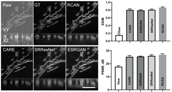
Excited to introduce #3DRCAN for denoising, super resolution and expansion microscopy. Super proud of this colab with Hari Shroff, Jiji Chen et al.
@NIBIBgov @arpcomplex
Preprint: biorxiv.org/content/10.110…
Code: github.com/AiviaCommunity…
#microscopy #aimicroscopy #aivia
1/17
@NIBIBgov @arpcomplex
Preprint: biorxiv.org/content/10.110…
Code: github.com/AiviaCommunity…
#microscopy #aimicroscopy #aivia
1/17
"Three-dimensional residual channel attention networks denoise and sharpen fluorescence microscopy image volumes"
J Chen, H Sasaki, H Lai, Y Su, J Liu, Y Wu, A Zhovmer, C Combs, I Rey-Suarez, H Chang, C Huang, X Li, M Guo, S Nizambad, A Upadhyaya, J Lee, L Lucas, H Shroff.
2/17
J Chen, H Sasaki, H Lai, Y Su, J Liu, Y Wu, A Zhovmer, C Combs, I Rey-Suarez, H Chang, C Huang, X Li, M Guo, S Nizambad, A Upadhyaya, J Lee, L Lucas, H Shroff.
2/17
We are particularly interested in characterizing the limits of #deeplearning based approaches to image restoration. You will find several experiments comparing several best-in-class approaches: #3DRCAN, #CARE, #SRResNet and #ESRGAN.
#microscopy #aimicroscopy #iSIM
3/17
#microscopy #aimicroscopy #iSIM
3/17

Pra deixar o pessoal de laboratório feliz e as aulas EAD menos complicadas, segue em anexo:
🔬THREAD COM SIMULAÇÕES DE LABORATÓRIO VIRTUAL🔬
#BiomedTwitter #AcademicTwitter #science #biology
🔬THREAD COM SIMULAÇÕES DE LABORATÓRIO VIRTUAL🔬
#BiomedTwitter #AcademicTwitter #science #biology
Nessa thread você vai encontrar simulações de laboratório de diagnóstico, #microscopy, #immunology, #molecularbiology, #cellbiology e tudo que eu encontrar. Dá uma olhada até o final pra ver se tem alguma simulação que te interessa! Vou atualizando aos poucos! :)
Antes de começar, gostaria de avisar que a maioria (talvez todas) das simulações são em inglês, infelizmente :(
🧬 Minha dica pra quem ainda não tem domínio da língua: ativa a tradução do google translate na página e vê se consegue entender o conteúdo da página. Talvez ajude!
🧬 Minha dica pra quem ainda não tem domínio da língua: ativa a tradução do google translate na página e vê se consegue entender o conteúdo da página. Talvez ajude!

#AfricanNeuroscience [Thread]
Ever wondered what #Neuroscience research looks like in #Africa? For 3 years, we have been working on profiling the neuroscience research landscape in the continent’s 54 countries. Very excited to share our latest work: biorxiv.org/content/10.110…
(1)
Ever wondered what #Neuroscience research looks like in #Africa? For 3 years, we have been working on profiling the neuroscience research landscape in the continent’s 54 countries. Very excited to share our latest work: biorxiv.org/content/10.110…
(1)
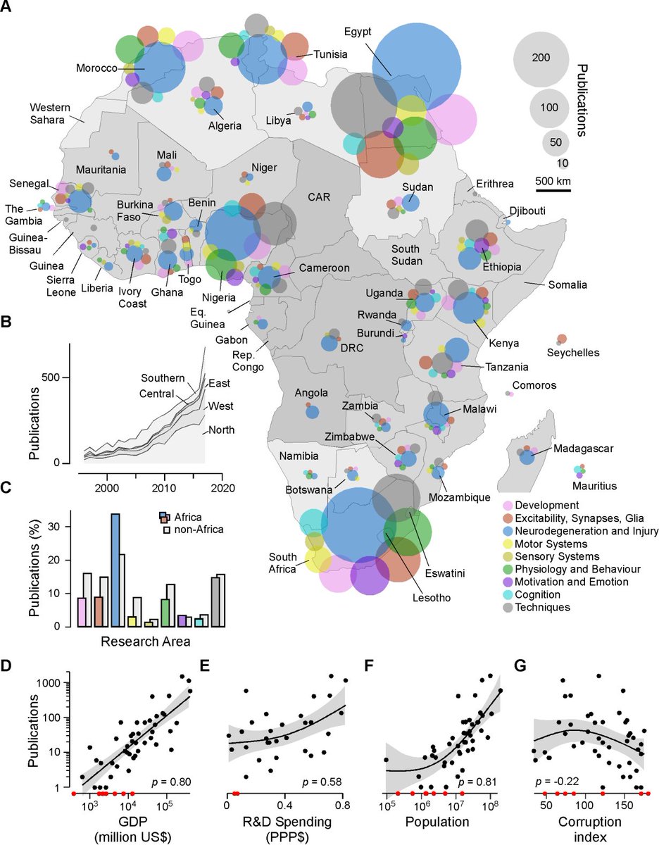
Why is this important?
#Africa has real potential in #Neuroscience, but needs help to rise from its current position! To do this, we need accurate data that reflects the heterogeneity of research across the continent's 54 countries.
(2)
#Africa has real potential in #Neuroscience, but needs help to rise from its current position! To do this, we need accurate data that reflects the heterogeneity of research across the continent's 54 countries.
(2)
Such data is lacking, as previous estimates about research outputs from #Africa are mostly inaccurate, partly because it is difficult to tell whether the research was, in fact, conducted within Africa or outside the continent in collaboration with #Africanscientists
(3)
(3)

Good news! This new human #tumor microbiome study is excellent. They used hundreds of negative controls for contamination + an extensive combo of methods to identify #bacteria in 7 #cancer types (lung, ovary, pancreas, melanoma, bone, brain): science.sciencemag.org/content/368/64…

2/ Bacterial LPS + 16SrRNA were frequently detected in all tumor types, w/ #breast cancer harboring a particularly rich and diverse #microbiome. The image below shows LPS/16SrRNA in breast cancer cells. Bacteria were also found in normal breast samples from healthy subjects


We found unique breast immune cells in the duct walls – ductal macrophages. They remove dying cells, help remodelling + are key players in cancer.
How did we get from this first sighting to finding their identity and function? Follow this thread!
bit.ly/35o2XSj
1/n
How did we get from this first sighting to finding their identity and function? Follow this thread!
bit.ly/35o2XSj
1/n
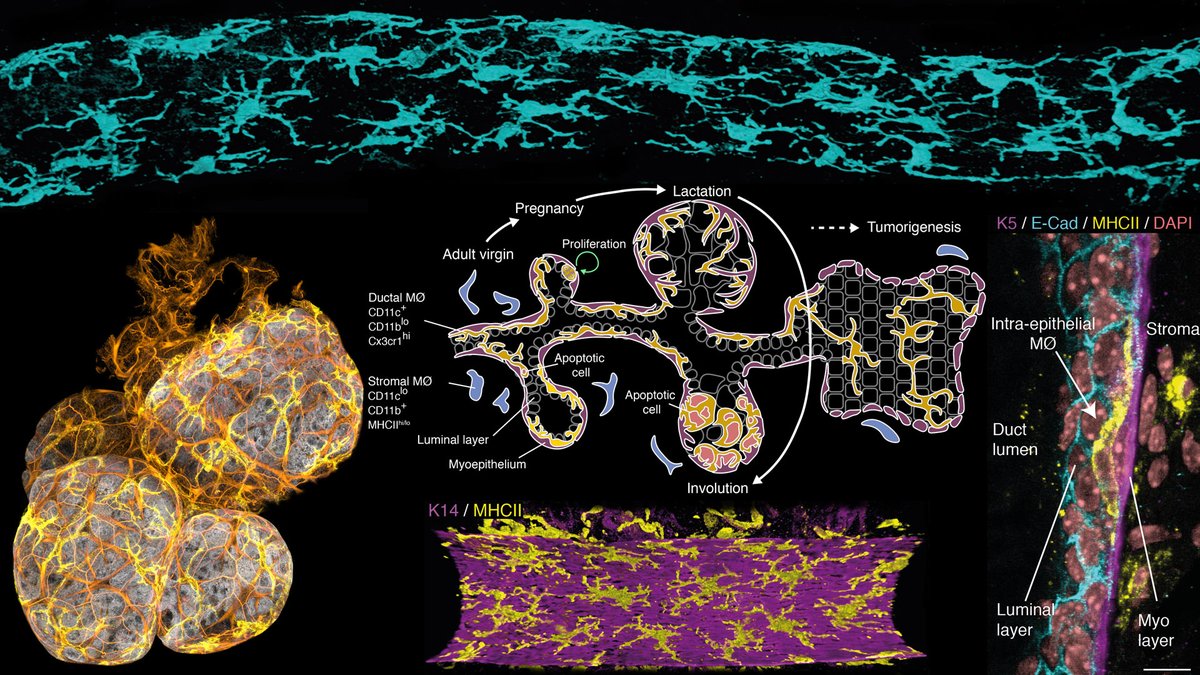
The breast contains mammary ducts surrounded by fat, blood vessels, immune + other cells.
We use 3D imaging to investigate the complex relationships b/w these cells to better understand disease.
#microscopy #mammarygland
2/n
We use 3D imaging to investigate the complex relationships b/w these cells to better understand disease.
#microscopy #mammarygland
2/n
The ducts form a tree that branches through the breast and blooms and recedes in pregnancy and weaning. These are precancerous mouse ducts, alveoli in lactation and a summary of development in mice.
3/n
3/n


@3dtrevor @Mariposa_Rising @MascalchiP and the wider team are running another well attended hands on online #bioimageanalysis workshop. Today about 3d segmentation and tracking! See more: drvtechnologies.com/aivia-online-w… #aivia #aimicroscopy #Microscopy

Uses the freely available #aiviaweb.
Setting ROIs to get quick previews.

The #OSABiophotonics20 (20-23 of April) congress has been moved to an 100% online format due to #covid19. This event is packed with great (microscopy and image analysis) presentations and is FREE to attend! Register here: osa.org/en-us/meetings… 1/3
#aimicroscopy #biophotonics
#aimicroscopy #biophotonics

My talk "AI for Microscopy Applications|The Era of Implementation", highlights 2+ years of R&D (colab w/ Hari Shroff et al.), and is influenced by @kaifulee @EricTopol @loicaroyer @martweig @florianjug @TheGeneMyers @uschmidt83 @PavelTomancak @HenriquesLab #OSABiophotonics20 2/3

KUDOS to @OpticalSociety @OSAPublishing and organizers for quickly moving the event to "online" and for making it free to participate. Also, I thank the organizers for the invite to present our work - MUCH appreciated. 3/3
#communityimpact #OpenScience #aimicroscopy #microscopy
#communityimpact #OpenScience #aimicroscopy #microscopy
My talk on #AIMicroscopy as presented at the "NIH AI Imaging Workshops".
Intro/history of AI, apps in #microscopy, intro to #deeplearning, and some of our recent research results into image restoration (in colab with Hari Shroff et al).
Organized by: @NCIDataSci @NIH @NIBIBgov
Intro/history of AI, apps in #microscopy, intro to #deeplearning, and some of our recent research results into image restoration (in colab with Hari Shroff et al).
Organized by: @NCIDataSci @NIH @NIBIBgov
The video of my part starts from 1:00:45.
Before that is a talk from Dr. Matt Guay from NIBIB (I recommend it too!). "An Introduction to Content-Aware Computation for Optical Microscopy" #CARE @loicaroyer @florianjug @uschmidt83 @martweig
Before that is a talk from Dr. Matt Guay from NIBIB (I recommend it too!). "An Introduction to Content-Aware Computation for Optical Microscopy" #CARE @loicaroyer @florianjug @uschmidt83 @martweig
We will start to roll out #AiviaWeb at no cost to use, tomorrow. All #aivia functionality accessible via a web browser. This is part of our #covid19 mitigation plan - more actions soon so you can continue to be productive while #wfh. #bioimageanalyis #aiviacommunity #microscopy

Once you are signed up we will reach out to you with all the details including usage and booking guidelines. A webpage dedicated to our covid19 / wfh mitigation plan will be up very soon too. Many more resources are in the pipeline.

*** 1/9 ***
Today is HIS-TO-RI-CAL about Aivia!!!
We are releasing a free to use software called Aivia Community
It’s meant to be a collaborative tool, allowing you to…
(see next tweets)
#Microscopy #ImageAnalysis #bioimageanalysis #3Drendering
Today is HIS-TO-RI-CAL about Aivia!!!
We are releasing a free to use software called Aivia Community
It’s meant to be a collaborative tool, allowing you to…
(see next tweets)
#Microscopy #ImageAnalysis #bioimageanalysis #3Drendering

*** 2/9 ***
Open and interactively explore 2-to-5D microscopy data sets
(tested up to 3 TB files)
Open and interactively explore 2-to-5D microscopy data sets
(tested up to 3 TB files)
*** 3/9 ***
Create attractive visual outputs (such as snapshots, video animation) from:
•raw microscopy images
•OR data sets previously analyzed with Aivia (with 3D surfaces, traced neurons for instance)
#3Drendering #Animations #VideoExport #Snapshots #3Dsurfaces
Create attractive visual outputs (such as snapshots, video animation) from:
•raw microscopy images
•OR data sets previously analyzed with Aivia (with 3D surfaces, traced neurons for instance)
#3Drendering #Animations #VideoExport #Snapshots #3Dsurfaces

1/n - Did you know developing #sperm cells are interconnected by cytoplasmic bridges until released from the testis? Just check out this awesome electron #microscopy 🔬image (by the late Don Fawcett) of two neighboring sperm cells. Note the arrow connecting them 🧐
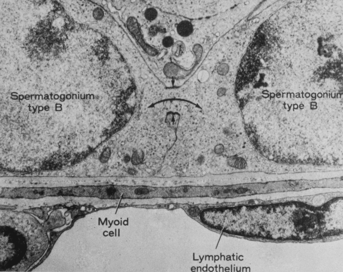
2/n- An intriguing property of these bridges is that the proteins that form them are also involved in the regulation of meiotic chromsomes 🤔 Topoisomerase 2 is a recently identified example 👇academic.oup.com/biolreprod/art…
3/3 - This suggests there's something more to these bridges than just being channels between neighboring cells... If you are curious about these intriguing structures stay tuned 📻 for future developments 🤓
1/ At #ASCBEMBO19 we will present five posters and a tech talk on #aimicroscopy and #CLEM. Details here: drvtechnologies.com/ascbembo2019
Many thanks to our collaborators Hari Shroff, Jiji Chen, @huzhao4, Rachel Wong, Chris Combs, @3i_inc.
#microscopy #imageanalysis #Aivia #ASCBEMBO2019
Many thanks to our collaborators Hari Shroff, Jiji Chen, @huzhao4, Rachel Wong, Chris Combs, @3i_inc.
#microscopy #imageanalysis #Aivia #ASCBEMBO2019

2/ P35/B36 Universal EM Connectomic analyses by Deep Learning Powered App‐matching Image Conversion
drvtechnologies.com/post/ascb-embo…
With Prof. Rachel Wong and @Sallywing
#ASCBEMBO19 #AImicroscopy #Aivia #AiviaCloud #imageanalysis #microscopy #CellBiology #machinelearning #drawacell
drvtechnologies.com/post/ascb-embo…
With Prof. Rachel Wong and @Sallywing
#ASCBEMBO19 #AImicroscopy #Aivia #AiviaCloud #imageanalysis #microscopy #CellBiology #machinelearning #drawacell

3/ P57/B58 GPU‐accelerated Machine Learning‐powered 3d Image Segmentation at Scale
Details: drvtechnologies.com/post/ascb-embo…
#ASCBEMBO19 #AImicroscopy #Aivia #AiviaCloud #imageanalysis #microscopy #CellBiology #machinelearning #drawacell #pixelclassifier #imagesegmentation
Details: drvtechnologies.com/post/ascb-embo…
#ASCBEMBO19 #AImicroscopy #Aivia #AiviaCloud #imageanalysis #microscopy #CellBiology #machinelearning #drawacell #pixelclassifier #imagesegmentation


How do you make nuclear pores away from the nucleus?
Our paper online today bit.ly/AL-NPC
@embl @a_schw @SchwabYannick @CellCellPress #development #Nuclearpores #microscopy
https://t.co/f4Xq1Vsk31
1/11 [a thread]
Our paper online today bit.ly/AL-NPC
@embl @a_schw @SchwabYannick @CellCellPress #development #Nuclearpores #microscopy
https://t.co/f4Xq1Vsk31
1/11 [a thread]
Background: Mothers stockpile nuclear pores (NPCs) in the cytoplasm of their eggs to enable embryonic cell cycles that are too fast for conventional NPC assembly (bit.ly/ALembryo). These are arranged in stacked ER sheets called Annulate Lamellae (AL, see 4/11).
2/11
2/11
These are Drosophila egg chambers; where the growing oocyte is transcriptionally silent, while so-called nurse cells produce the material for its development and transport it to the front. We wanted to see if this includes AL and whether they accumulate over time.
3/11
3/11

