Discover and read the best of Twitter Threads about #neuroanatomy
Most recents (23)

A fascinating symptom in #neurology is #Alien Hand syndrome
If anyone has seen @StanleyKubrick ‘s great film Dr. Strangelove, you might have wondered what’s wrong with the titular character
His hand has a #mind of its own!!!
#cinema #neurotwitter #MedTwitter
If anyone has seen @StanleyKubrick ‘s great film Dr. Strangelove, you might have wondered what’s wrong with the titular character
His hand has a #mind of its own!!!
#cinema #neurotwitter #MedTwitter
Simplifying , for carrying out bimanual planned movements, we need a Supplementary Motor Cortex (SMA) (left frontal lobe). This communicates to the opposite side via the corpus callosum. Lesions in any will impair proper bimanual function and cause an “alien limb”#neuroanatomy
Frontal variant (left sma lesion) : Patient will have impulsive groping and difficulty releasing objects
Affects the dominant hand
Can be due to a #stroke, #degeneration
My patient impulsively grabs my hand (with his right hand) despite me telling him not to!
#neurotwitter
Affects the dominant hand
Can be due to a #stroke, #degeneration
My patient impulsively grabs my hand (with his right hand) despite me telling him not to!
#neurotwitter

4⃣ core brain networks (there are more): expanding the localizationist approach 🧠 🥅🕸️
#Neuro #Neuroanatomy #Neurology #EndNeurophobia
#Thread
1/🧵
#Neuro #Neuroanatomy #Neurology #EndNeurophobia
#Thread
1/🧵
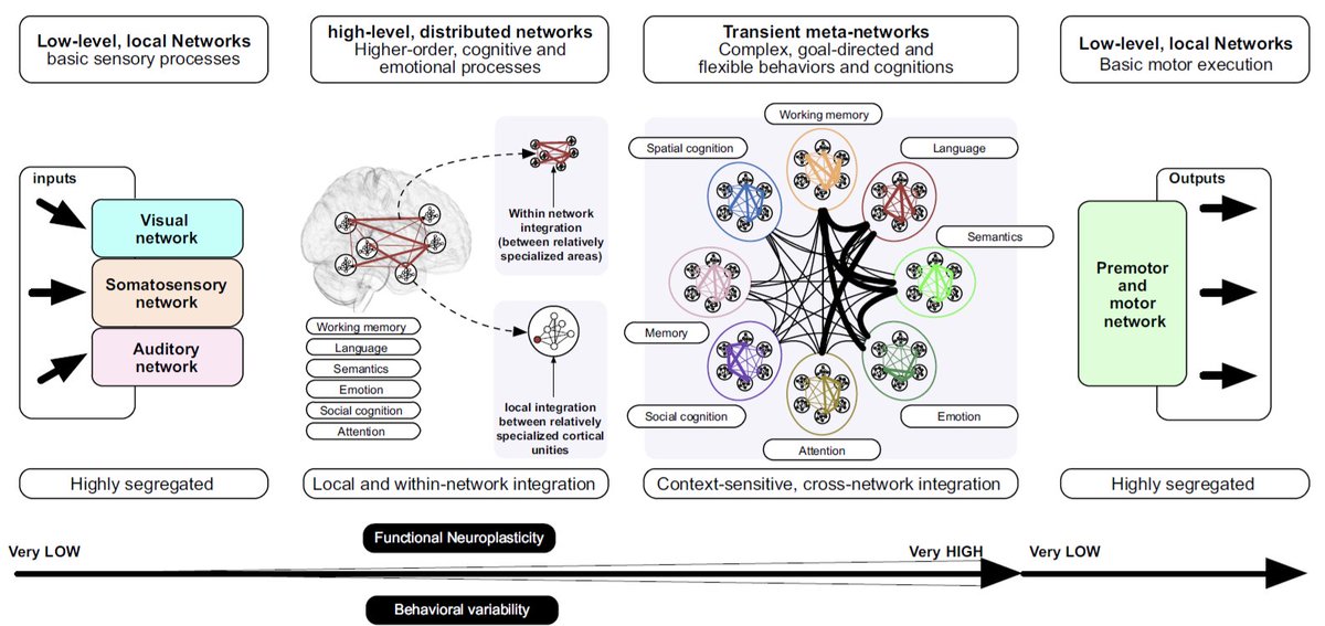
1⃣ Default mode network
🥇 Endogenously mediated activities at rest
🥈 Self-referential and social cognitive processes,
🚫Not active during external goal-oriented processes
2/🧵
🥇 Endogenously mediated activities at rest
🥈 Self-referential and social cognitive processes,
🚫Not active during external goal-oriented processes
2/🧵
1⃣ Default mode network
Components
💠 Posterior cingulate cortex
💠 Medial prefrontal cortex
💠 Precuneus
💠 Inferior parietal and medial temporal cortices
3/🧵
Components
💠 Posterior cingulate cortex
💠 Medial prefrontal cortex
💠 Precuneus
💠 Inferior parietal and medial temporal cortices
3/🧵

HYPOTHALAMUS (HT)🧵-the control center of circadian rhythm, fatigue/wakefulness, hunger/satiety, sex drive, thirst/BP—and the command post for endocrine control via the HT-pituitary axis.
#meded #neuroradiology #neuroscience #radiology #neurology #neurosurgery #neuroanatomy
1/28
#meded #neuroradiology #neuroscience #radiology #neurology #neurosurgery #neuroanatomy
1/28
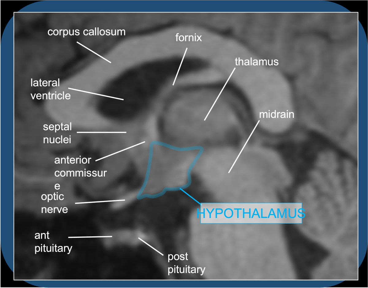
Quiz A: Histamine activity in the brain/brainstem contributes to alertness/wakefulness–hence: sleepy effects of benadryl. Histaminergic neurosecretory cell bodies are exclusively in the tuberomammillary nucleus of the TUBER CINEREUM (HT floor), with widespread projections.
2/28
2/28

The HT is a mysterious and complex—I get confused sighs from medical students when the topic arises—the small almond-sized morsel is the control center for endocrine/hormone regulation, and homeostasis of food/water consumption, sleep, BP, sex/attachment. The HT is boss!
3/28
3/28
Time to review one of my favorite topics in Neuroanatomy. The thalamus: from myth to neurology. 🧠🍏🚣
#Neurotwitter #Neurology #Neuroanatomy #Endneurophobia.
1/🧵
#Neurotwitter #Neurology #Neuroanatomy #Endneurophobia.
1/🧵
First of all a brief disclaimer.
The objective of the following thread is to illustrate in a practical and easy way the thalamic anatomy. Details on the newest advances in classification and function are outside of the main objective. 🧑🏫🏥⚕️
2/🧵
The objective of the following thread is to illustrate in a practical and easy way the thalamic anatomy. Details on the newest advances in classification and function are outside of the main objective. 🧑🏫🏥⚕️
2/🧵
The word "Thalamus" comes from the greek word "θάλαμος" which means chamber. It was used to describe the main bedroom (mostly the bridal room) in a house in the ancient greece. 🛏️
3/🧵
3/🧵

Hey #Medtwitter & #Neurotwitter With 2 simple rules you can learn the #basalganglia direct and indirect pathways for the LAST TIME and actually remember them! 🤯Here’s how! 🧵 :
#MedEd #medicalstudent #Neurology #Neurologyresident #Movementdisorders #Neuroscience #Neuroanatomy
#MedEd #medicalstudent #Neurology #Neurologyresident #Movementdisorders #Neuroscience #Neuroanatomy
Rule 1: The “OG” basal ganglia (striatum and globus pallidi) use GABA ➖as their neurotransmitter. Everything else uses glutamate ➕.
Rule 2: Once you know Rule 1, all you have to do is memorize the order of the structures:
Direct pathway: CS___GiT
Indirect pathway: CS GeS GiT
Rule 2: Once you know Rule 1, all you have to do is memorize the order of the structures:
Direct pathway: CS___GiT
Indirect pathway: CS GeS GiT
Both pathways start with
cortex (C)>striatum (S)
and end with globus pallidus internus (Gi)>Thalamus (T).
For the indirect, just add GeS (Globus pallidus externus>STN) in the middle!
cortex (C)>striatum (S)
and end with globus pallidus internus (Gi)>Thalamus (T).
For the indirect, just add GeS (Globus pallidus externus>STN) in the middle!
Pterygopalatine fossa🧵- inspired by ?s from med students in neuroanatomy lab & a resident w/ case of perineural tumor spread on same day! #meded #FOAMrad #medtwitter #medstudents #radiology #neurorad #HNrad #radres #neurology #ENT #temporalbone #neurosurgery #neuroanatomy
1/22
1/22
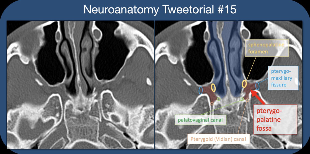
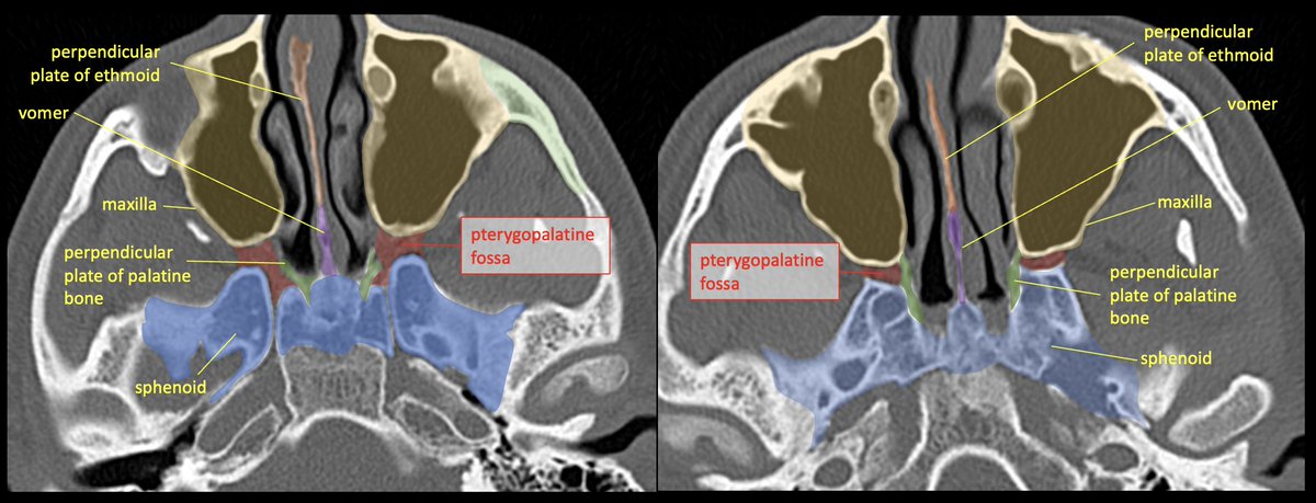
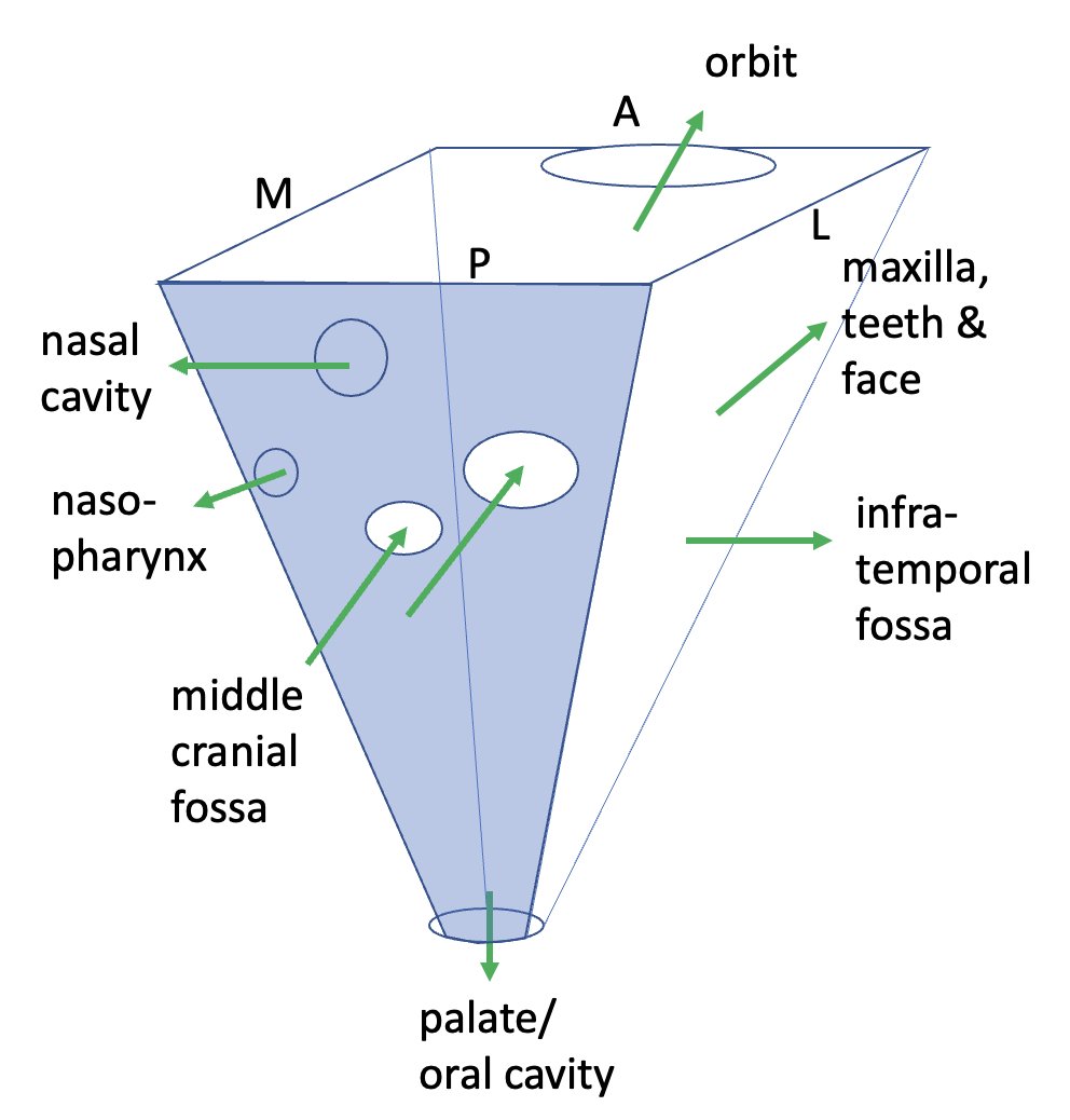
The PTERYGOPALATINE FOSSA (PPF) is a space deep in the face/skull base, bordered anteriorly by the maxilla (max sinus), posteriorly by the pterygoid base of the sphenoid, and medially by the perpendicular plate of the palatine bone. 2/22
It’s best to think of the PPF as a crossroads/intersection. Think about the roads that lead to and from it, and the cast of characters that pass through.
Some like to simplify/visualize the PPF as a cube or inverted pyramid.
3/22
Some like to simplify/visualize the PPF as a cube or inverted pyramid.
3/22

Learning brainstem #neuroanatomy would be nearly impossible without the rule of 4s. I'm going to pass this along for anyone trying to learn #neurology, as this might be the single most powerful mnemonic I know of.
I present to you, The Rule of 4s: a #MedEd #tweetorial
(1/10)
I present to you, The Rule of 4s: a #MedEd #tweetorial
(1/10)
4 cranial nerves per brainstem level
- Midbrain: 1-4
- Pons: 5-8
- Medulla: 9-12
* CNs 1 and 11 do not connect with the brainstem
* CN5 enters in the pons, but has nuclei at all three levels
(2/10)
- Midbrain: 1-4
- Pons: 5-8
- Medulla: 9-12
* CNs 1 and 11 do not connect with the brainstem
* CN5 enters in the pons, but has nuclei at all three levels
(2/10)
4 lateral (side) structures, all with an "S":
- Spinocerebellar pathway
- Spinothalamic pathway
- Sensory nucleus of CN5
- Sympathetic pathway
(3/10)
- Spinocerebellar pathway
- Spinothalamic pathway
- Sensory nucleus of CN5
- Sympathetic pathway
(3/10)
Neuroanatomy TOTD #15🧵
The inner ear #tweetorial--it packs a large functional punch for its small size-strap in!
#meded #FOAMed #FOAMrad #medtwitter #medstudents #radiology #neurorad #HNrad #radres #neurology #ENT #temporalbone #neurosurgery #neuroanatomy #neuroanatomyTOTD
1/24
The inner ear #tweetorial--it packs a large functional punch for its small size-strap in!
#meded #FOAMed #FOAMrad #medtwitter #medstudents #radiology #neurorad #HNrad #radres #neurology #ENT #temporalbone #neurosurgery #neuroanatomy #neuroanatomyTOTD
1/24
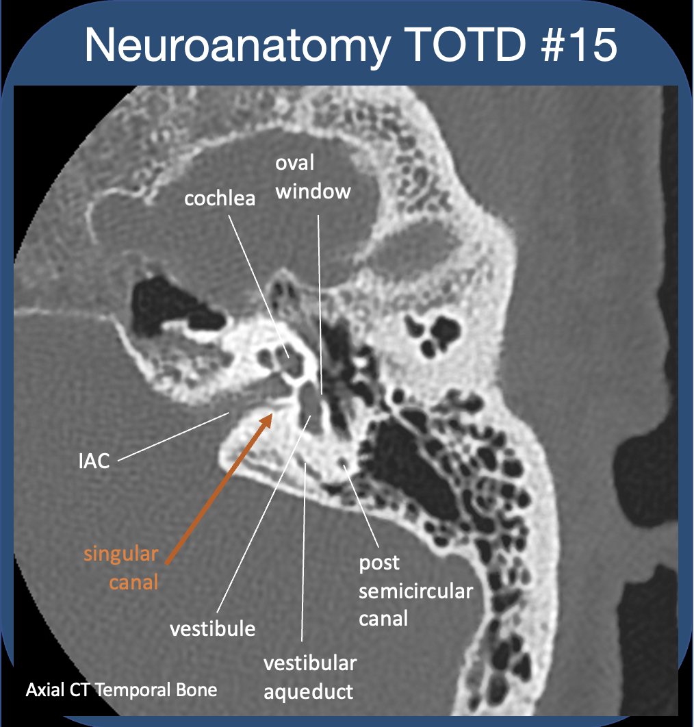
To evaluate the t-bone, best to compartmentalize--external/middle/inner ear (IE). See previous #tweetorial of the ME. The IE is difficult as most structures are obliquely oriented (at different obliquities!)-and can be hard to see on standard views. 2/24

IE communicates with ME via oval&round windows (which allow for transmission&dissipation of sonic vibrations). IE houses sensory organs for hearing/balance/sensing motion. The cochlear&vestibular nerves (CNVIII) transmit signals to the brain via the int auditory canal (IAC). 3/24

Neuroanatomy TOTD #14🧵
Got some requests to do one of the trickiest areas of human anatomy, the #temporalbone. So many named structures! #meded #FOAMed #FOAMrad #medtwitter #medstudents #radiology #neurorad #radres #neurosurgery #neuroanatomy #ENT #otolaryngology
1/21
Got some requests to do one of the trickiest areas of human anatomy, the #temporalbone. So many named structures! #meded #FOAMed #FOAMrad #medtwitter #medstudents #radiology #neurorad #radres #neurosurgery #neuroanatomy #ENT #otolaryngology
1/21
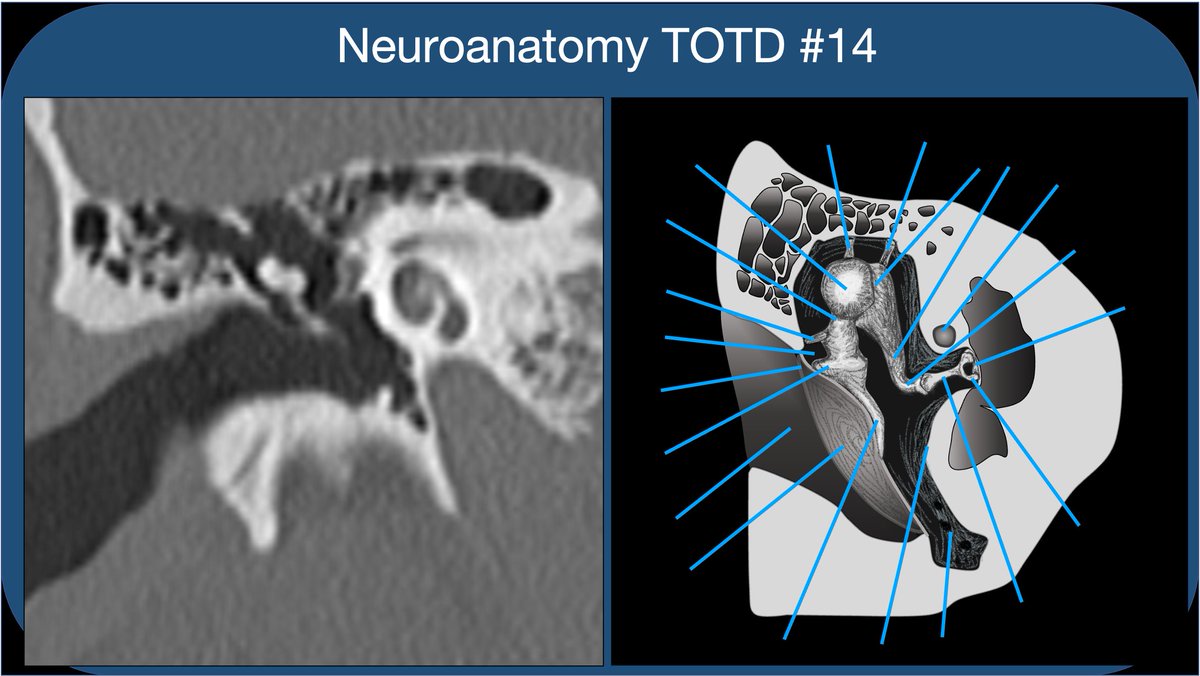
Whether learning t-bone anatomy as a medical student or evaluating a CT of the t-bone as a radres, it’s best to compartmentalize into external, middle, and inner ear. This thread🧵is on the middle ear: Inner ear to follow, some day:)
2/21
2/21

The external ear extends from the external meatus to the TM. The TM should be thin and *almost* imperceptible on CT. Thickened and retracted TM suggests prior pathology (usually otitis media) and scarring.
3/21
3/21

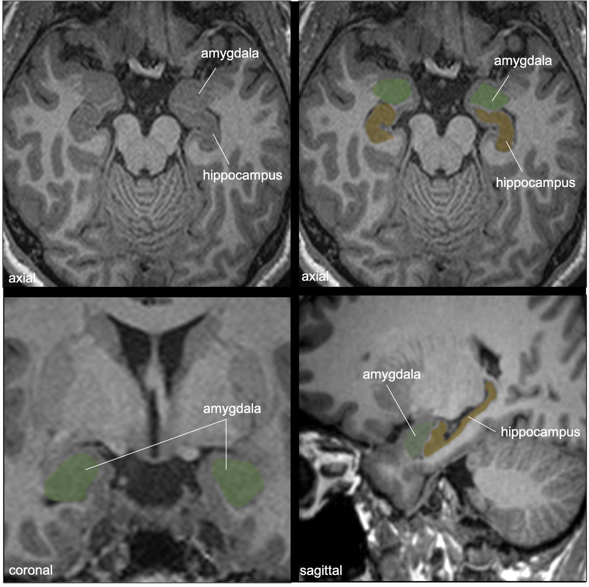
Neuroanatomy TOTD #12
The green structure is the amygdala (amygdaloid body) and the yellow structure is the stria terminalis (ST).
1/18
#meded #FOAMed #FOAMrad #medtwitter #medstudents #radiology #neurorad #radres #neurology #neurosurgery #neuroanatomy #neuroanatomyTOTD
The green structure is the amygdala (amygdaloid body) and the yellow structure is the stria terminalis (ST).
1/18
#meded #FOAMed #FOAMrad #medtwitter #medstudents #radiology #neurorad #radres #neurology #neurosurgery #neuroanatomy #neuroanatomyTOTD

Time for a deep dive into limbic networks. Bear with me—this is a fun subject. I got carried away preparing slides—it’s hard to know when to stop!
2/18
2/18
The amygdala is an almond-shaped collection of gray nuclei/subnuclei deep to the uncus and ant to hippocampus in the med temporal lobe. Involved in multiple functions: memory modulation, emotional learning and responses, +important connections w/ the olfactory bulb/cortex.
3/18
3/18
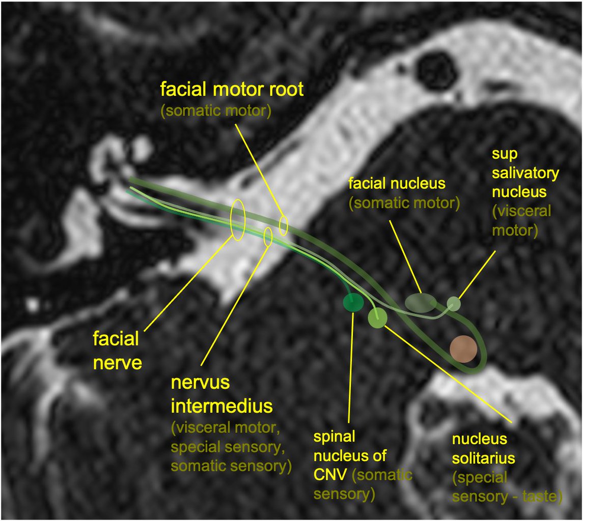
Neuroanatomy TOTD #11🧵
➡️intracanalicular facial nerve. IAC cross section➡️4 nerves; ant-sup➡️facial, ant-inf➡️cochlear (7-up/coke-down). Post nerves➡️sup&inf vestibular #medtwitter #meded #FOAMed #FOAMrad #neurorad #radiology #radres #neurology #neurosurgery #neuroanatomy
1/11
➡️intracanalicular facial nerve. IAC cross section➡️4 nerves; ant-sup➡️facial, ant-inf➡️cochlear (7-up/coke-down). Post nerves➡️sup&inf vestibular #medtwitter #meded #FOAMed #FOAMrad #neurorad #radiology #radres #neurology #neurosurgery #neuroanatomy
1/11

Facial motor nucleus is in the pontine tegmentum; axons loop dorsally around the abducens nucleus, then course anterolaterally, exiting the brainstem at the CPA.
2/11
2/11
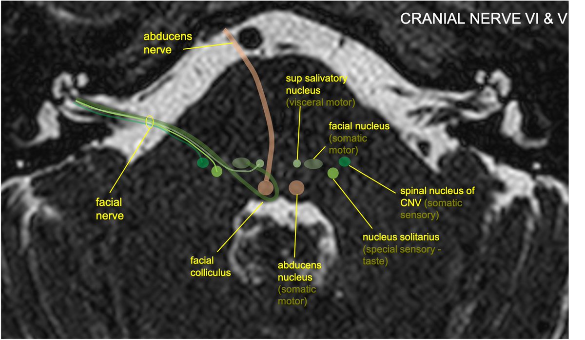
The nervus intermedius (NI) is composed of preganglionic parasympathetic fibers w/cell bodies in the sup salivatory nucleus, taste fibers w/cell bodies in the nucleus solitarius, and somatic sensory input from EAC/external ear, w/cell bodies in the spinal nucleus of CNV.
3/11
3/11
Neuroanatomy TOTD #10🧵
1/5 Small gray matter structure at the junction of the thalamus and midbrain is the subthalamic nucleus (STN).
#meded #FOAMed #FOAMrad #medtwitter #medstudents #radiology #neurorad #radres #neurology #neurosurgery #neuroanatomy #neuroanatomyTOTD
1/5 Small gray matter structure at the junction of the thalamus and midbrain is the subthalamic nucleus (STN).
#meded #FOAMed #FOAMrad #medtwitter #medstudents #radiology #neurorad #radres #neurology #neurosurgery #neuroanatomy #neuroanatomyTOTD

2/5...The STN is functionally a node within the basal ganglia (BG) INDIRECT LOOP. STN contains excitatory glutaminergic neurons➞output to the GABA neurons of GPi, which in turn have inhibitory effect on thalamic outputs to the motor cortex.

3/5...Loss of nigrostriatal input in Parkinsons Dz➞increased inhibitory output from GPi➞decreased thalamic stimulation of the motor cortex (through both direct and indirect loop circuits). Makes sense that DBS treatments were initially directed at disrupting activity in GPi.

Neuroanatomy TOTD #9🧵
1/6 The trigeminal n. courses anteriorly➡️prepontine cistern➡️into Meckel’s cave (green), which lies at the medial floor of middle cranial fossa at the petrous apex.
#meded #FOAMed #FOAMrad #neurorad #neurology #neurosurgery #neuroanatomy #neuroanatomyTOTD
1/6 The trigeminal n. courses anteriorly➡️prepontine cistern➡️into Meckel’s cave (green), which lies at the medial floor of middle cranial fossa at the petrous apex.
#meded #FOAMed #FOAMrad #neurorad #neurology #neurosurgery #neuroanatomy #neuroanatomyTOTD

2/6...Meckel’s cave (MC) is open to the subarachnoid space at its posterior margin (and is therefore filled with CSF). The trigeminal (Gasserion) ganglion lives in MC. Superiomedial to MC (and sharing a dural border), is the cavernous sinus. #radres #neurorad

3/6...Coursing anteriorly, the V1 and V2 branches of CNV exit MC to travel within the lateral wall of the cavernous sinus. V3 courses inferiorly to exit the middle cranial fossa through foramen ovale, without involving the cavernous sinus.

Neuroanatomy TOTD #8🧵
1/8 MRI shows the hippocampal formation; the hippocampus(HC) is a ridge of archicortex gray matter at floor of lat ventricle, in the med temp lobe #FOAMed #FOAMrad #radres #neurorad #neuroscience #medtwitter #radiology #neurology #neurosurgery #neuroanatomy
1/8 MRI shows the hippocampal formation; the hippocampus(HC) is a ridge of archicortex gray matter at floor of lat ventricle, in the med temp lobe #FOAMed #FOAMrad #radres #neurorad #neuroscience #medtwitter #radiology #neurology #neurosurgery #neuroanatomy

2/8..The HC: complex structure of limbic system; encodes memories from short->long term (also involved in pattern recognition,memory encoding & association, working memory,spatial nav, emotional behavior,awareness of conscious knowledge). Am I missing anything neuroscientists?
3/8...Anatomically: the HC is made of the cornu ammonis (CA) and dentate gyrus (DG); hippocampal formation also includes the subiculum and entorhinal cortex (EC). Can also be divided into head/body/tail. Supplied primarily by the PCA, and variably by anterior choroidal artery.

Neuroanatomy TOTD #7🧵
1/7 The cavernous sinuses (CS) are outlined on either side of the sella. The ICA & cranial nerves 3,4,6,V1,V2 travel through the CS.
#meded #FOAMed #FOAMrad #radres #neurorad #medtwitter #radiology #neurology #neurosurgery #neuroanatomy #neuroanatomyTOTD
1/7 The cavernous sinuses (CS) are outlined on either side of the sella. The ICA & cranial nerves 3,4,6,V1,V2 travel through the CS.
#meded #FOAMed #FOAMrad #radres #neurorad #medtwitter #radiology #neurology #neurosurgery #neuroanatomy #neuroanatomyTOTD

2/7...CN 3,6,V1 travel between two dural layers in the lateral wall of the CS. V2 often travels in the the inferolateral wall of the CS (sometimes inferior to the CS). Cranial nerve 6 floats freely in the CS (why CS pathology often selectively affects CN6).

3/7...Note that: postganglionic sympathetic inputs to the orbit (originating from the sup cervical ganglion) ascend with the ICA, branch off the ICA in the CS, and then join branches of CN3 (to sup tarsal muscle) and V1 (pupillary dilation).
Neuroanatomy TOTD #6
a) Ant choroidal artery infarct, involving the post limb of the internal capsule
b) Sup hypophyseal artery aneurysm—medially directed from the supraclinoid ICA
#meded #FOAMed #FOAMrad #radres #neurorad #radiology #neurosurgery #neuroanatomy #neuroanatomyTOTD
a) Ant choroidal artery infarct, involving the post limb of the internal capsule
b) Sup hypophyseal artery aneurysm—medially directed from the supraclinoid ICA
#meded #FOAMed #FOAMrad #radres #neurorad #radiology #neurosurgery #neuroanatomy #neuroanatomyTOTD


ICA segments: C1 (cervical) becomes C2 (petrous) in the carotid canal of the petrous bone. Becomes C3 (lacerum) as it exits the carotid canal above the foramen lacerum. Becomes C4 (cavernous segment above the petrolingual ligament through the cavernous sinus.

...C5 (clinoid) above proximal to the distal dural ring. C6 (ophthalmic) is truly intracranial. C7 (communicating) distal to the Pcomm. Alternative segmentation schemes include C1-C4 (cervical, petrous, cavernous, supraclinoid/terminal)

Neuroanatomy TOTD #5
The indicated bundle is the anterior commissure (AC), located at the ant border of the 3rd ventricle, at the sup margin of the lamina terminalis.
#meded #FOAMed #FOAMrad #radres #neurorad #medtwitter #radiology #neurosurgery #neuroanatomy #neuroanatomyTOTD
The indicated bundle is the anterior commissure (AC), located at the ant border of the 3rd ventricle, at the sup margin of the lamina terminalis.
#meded #FOAMed #FOAMrad #radres #neurorad #medtwitter #radiology #neurosurgery #neuroanatomy #neuroanatomyTOTD

The AC runs across the midline in front of the anterior columns of the fornix, behind the basal forebrain and beneath the anterior limb internal capsule and basal ganglia, surrounded by the bed nucleus of the stria terminalis.
The AC connects areas of the bilateral temporal poles and orbitofrontal cortex. Function is not entirely understood but it is thought to be important in the olfactory pathway and pain sensation, among other things.
Neuroanatomy TOTD #4
1/5 Answer: The orange structure is the cavernous sinus (CS), a paired dura-lined venous cavity on either side of the sella. The sinuses are split into numerous “caves” by fibrous septae (hence the name). #neuroanatomy #neurorad #medtwitter #neuroanatomyTOTD
1/5 Answer: The orange structure is the cavernous sinus (CS), a paired dura-lined venous cavity on either side of the sella. The sinuses are split into numerous “caves” by fibrous septae (hence the name). #neuroanatomy #neurorad #medtwitter #neuroanatomyTOTD
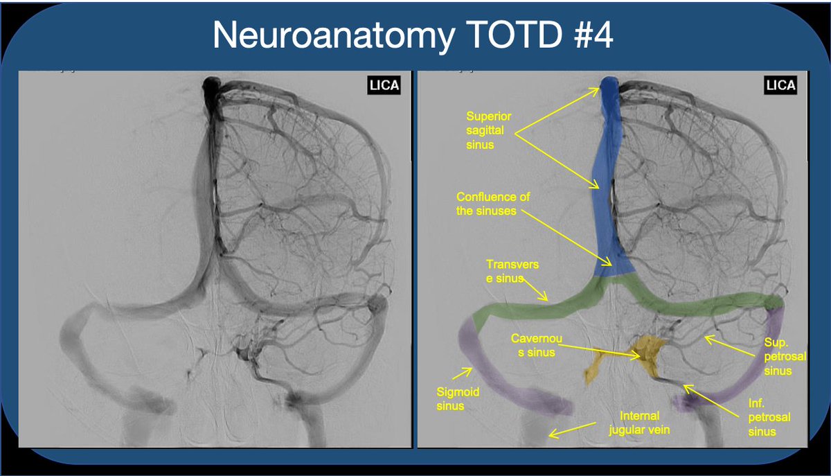
2/5 Note that the paired sinuses are often variably connected by “intercavernous sinuses”. While the CS is often taught for its relationship to the ICA and cranial nerves, I find that medical students and residents rarely understand the flow of blood through the sinus.
3/5 The CS receives blood from sup. and inf. ophthalmic veins draining the orbit--This is how a facial/orbital infection spreads intracranially to CS (classic #usmle ?). The sphenoparietal sinus as well as the superficial middle and inferieor cerebral veins also feed into the CS.

Neuroanatomy TOTD #3
1/6 The superior frontal sulcus courses in the AP direction, and terminates at the precentral sulcus posteriorly. The central sulcus is immediately posterior to the precentral, and the postcentral immediately posterior to the central.
#meded #FOAMed #FOAMrad
1/6 The superior frontal sulcus courses in the AP direction, and terminates at the precentral sulcus posteriorly. The central sulcus is immediately posterior to the precentral, and the postcentral immediately posterior to the central.
#meded #FOAMed #FOAMrad

2/6 "Upper T sign" or "L sign"
The SFS intersects the precentral s
Central sulcus is just posterior
The SFS intersects the precentral s
Central sulcus is just posterior

3/6 "Lower T sign" or "M sign"
The IFS terminates at the precentral s.
Central sulcus is just posterior
The IFS terminates at the precentral s.
Central sulcus is just posterior


Time for another #neuroanatomy #tweetorial #medstudent twitter!
By popular request, this one is On the VISUAL PATHWAY and localizing VISUAL FIELD DEFICITS!
cc @DxRxEdu @CPSolvers @CrystalYeoMDPhD @Tracey1milligan
1/
By popular request, this one is On the VISUAL PATHWAY and localizing VISUAL FIELD DEFICITS!
cc @DxRxEdu @CPSolvers @CrystalYeoMDPhD @Tracey1milligan
1/
But first, a moment of silence for #GeorgeFloyd #AhmaudArbery #BreonaTaylor
.
.
.
.
.
.
.
.
.
.
.
2/
.
.
.
.
.
.
.
.
.
.
.
2/
I’m grateful for all the ❤️ these #EndNeurophobia #tweetorials have received, but hope you’ll check out the rest of my feed too, which seeks to amplify the voices of those who I’m learning from in the path to being an antiracist ally.
3/
3/
Back with another #neurology #neuroanatomy #tweetorial.
This one goes out to all the #MedStudentTwitter studying
for the steps:
The BRAINSTEM and CRANIAL NERVES
#MedEd #NoMoreNeurophobia cc: @MadSattinJ @Tracey1milligan @StaceyLClardy @DxRxEdu @CPSolvers
This one goes out to all the #MedStudentTwitter studying
for the steps:
The BRAINSTEM and CRANIAL NERVES
#MedEd #NoMoreNeurophobia cc: @MadSattinJ @Tracey1milligan @StaceyLClardy @DxRxEdu @CPSolvers
This is often considered one of the most complex parts of neuroanatomy.
Let’s break it down:
First, the brainstem is divided into 3 levels, from superior to inferior:
MIDBRAIN
PONS
MEDULLA
These are MRI view so anterior on top, posterior on the bottom
Let’s break it down:
First, the brainstem is divided into 3 levels, from superior to inferior:
MIDBRAIN
PONS
MEDULLA
These are MRI view so anterior on top, posterior on the bottom

Midbrain= looks like Mickey Mouse
Pons=two huge cables connecting it to the cerebellum (middle cerebellar peduncles)
Medulla=looks almost like the spinal cord
Pons=two huge cables connecting it to the cerebellum (middle cerebellar peduncles)
Medulla=looks almost like the spinal cord

@DrNeuro123 @AaronLBerkowitz In muscular dystrophies with leg involvement, the calf muscles and leg muscles are affected first. Ext dig profundus (EDB) overcompensates for the foot drop caused by atrophy of the tibialis anterior (TA) so it hypertrophies as it tries desperately to lift the toes. (1)
@DrNeuro123 @AaronLBerkowitz In generalized neuropathies like CMT, the nerve dies backwards so by the time the nerve to the TA is affected, the nerve to the EDB has already been affected, so the EDB is atrophied just like the TA. So it’s a useful way to determine whether it’s a nerve or muscle problem. (2)
@DrNeuro123 @AaronLBerkowitz Peroneal neuropathies and L5/S1 radiculopathies are common causes of foot drop. When you examine foot drop, check foot inversion because that’s done by the tibialis posterior which is innervated by the tibial nerve. If that’s involved, it can’t be just the peroneal nerve (3)
I had no idea how popular #neuroanatomy #tweetorials would be! Thanks for the❤️!
Here's innervation of the leg/foot & approach to FOOT DROP
#MedEd #MedStudentTwitter
@CPSolvers @Tracey1milligan @MadSattinJ
@MedTweetorials @DxRxEdu @AANMember @StaceyLClardy @ContinuumAAN
1
Here's innervation of the leg/foot & approach to FOOT DROP
#MedEd #MedStudentTwitter
@CPSolvers @Tracey1milligan @MadSattinJ
@MedTweetorials @DxRxEdu @AANMember @StaceyLClardy @ContinuumAAN
1
Just like you can localize most peripheral upper extremity weakness by just knowing 5 nerves (see prior tweetorial), you can localize most peripheral lower extremity weakness by just knowing 5 nerves!
Just 5 nerves for each extremity- no neurophobia needed!
2
Just 5 nerves for each extremity- no neurophobia needed!
2
Which 5?
Femoral
Obturator
Sciatic
Peroneal
Tibial
And the sciatic is really just the peroneal and tibial bound together!
And the leg/foot are much easier than the hand because the foot does much less intricate movements than the hand!
3/
Femoral
Obturator
Sciatic
Peroneal
Tibial
And the sciatic is really just the peroneal and tibial bound together!
And the leg/foot are much easier than the hand because the foot does much less intricate movements than the hand!
3/
